Modern coating systems fulfill a variety of functions that go far beyond corrosion protection. The perceived value of various everyday objects, such as motor vehicles, household items or consumer electronics, is usually largelly defined by the visual appearance of their visible surfaces. The desire for a very high-quality visual appearance is the reason for the triumph of effect pigments.


If these effect pigments are embedded in complex polymer coating layers, a variety of high-gloss or sparkle effects can be achieved. In addition to the optical characteristics of effect pigments, functional properties such as electrical conductivity, reflectivity in the infrared range, laser markability or weather resistance are increasingly becoming the focus of development. In the Business Unit "Optical Materials and Technologies", nanostructure-based diagnostics methods have been used to examine effect pigments during development and production for more than 20 years.
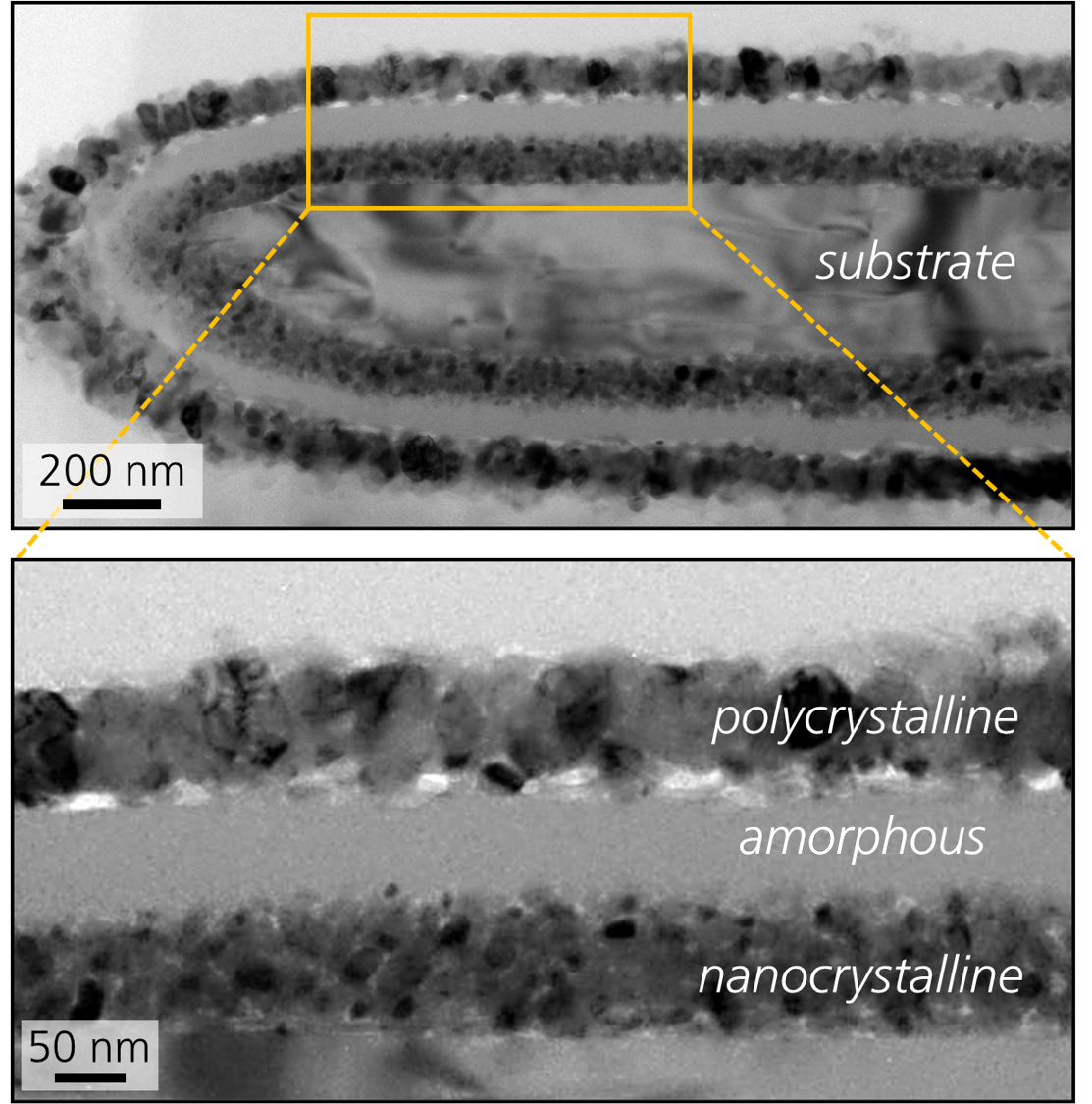
When designing effect pigments, interference layer systems are coated on a substrate particle, whose composition and sublayer thicknesses significantly define the optical appearance. These platelet-shaped particles typically have diameters in the range of a few micrometers, are a few 100 nm thick and are surrounded by layer systems consisting of individual layers of a few nanometers.
The following questions often arise during the development and production support of such materials:
- How can such small objects be prepared and then examined in cross-section without changing their initial state and introducing new artifacts?
- Which surface and interface roughness do the pigments have?
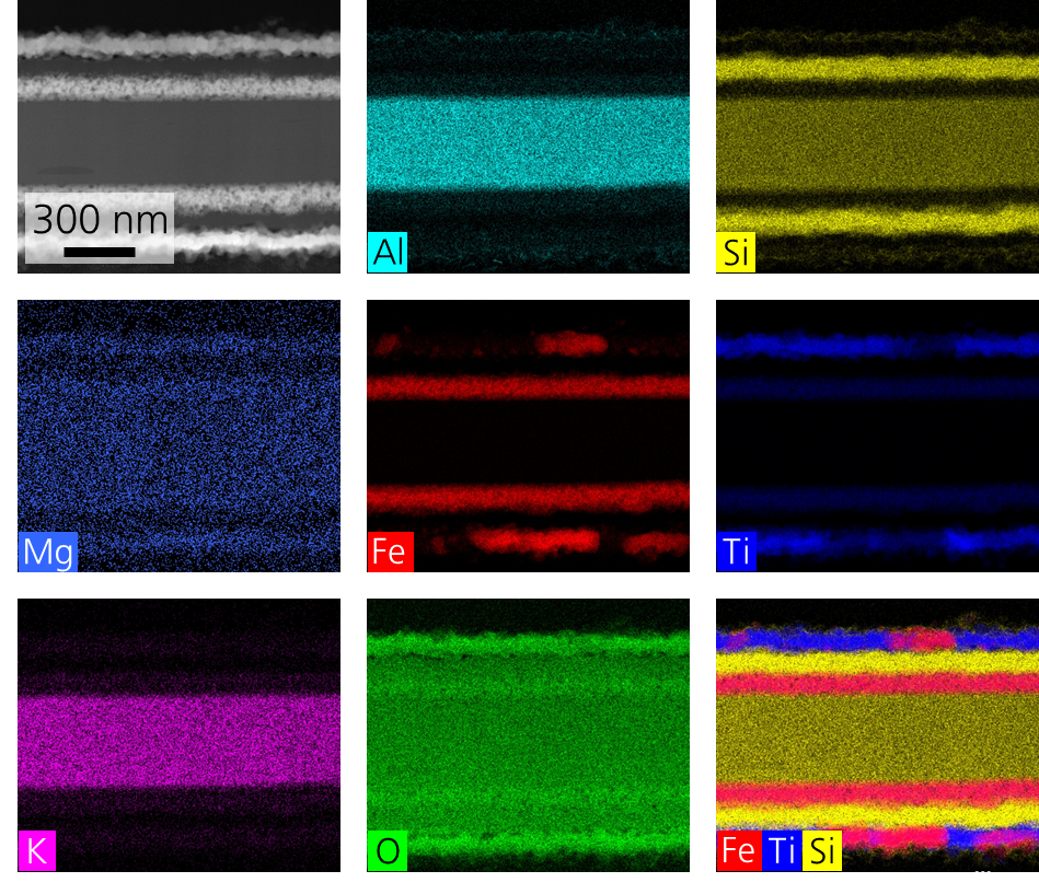
- How are the elements distributed in the effect pigments?
- Are the individual layers crystalline or amorphous and what layer thicknesses and homogeneity do they have?
- What grain sizes are present?
- Are there interactions between the layers? How well do the layers adhere to each other?
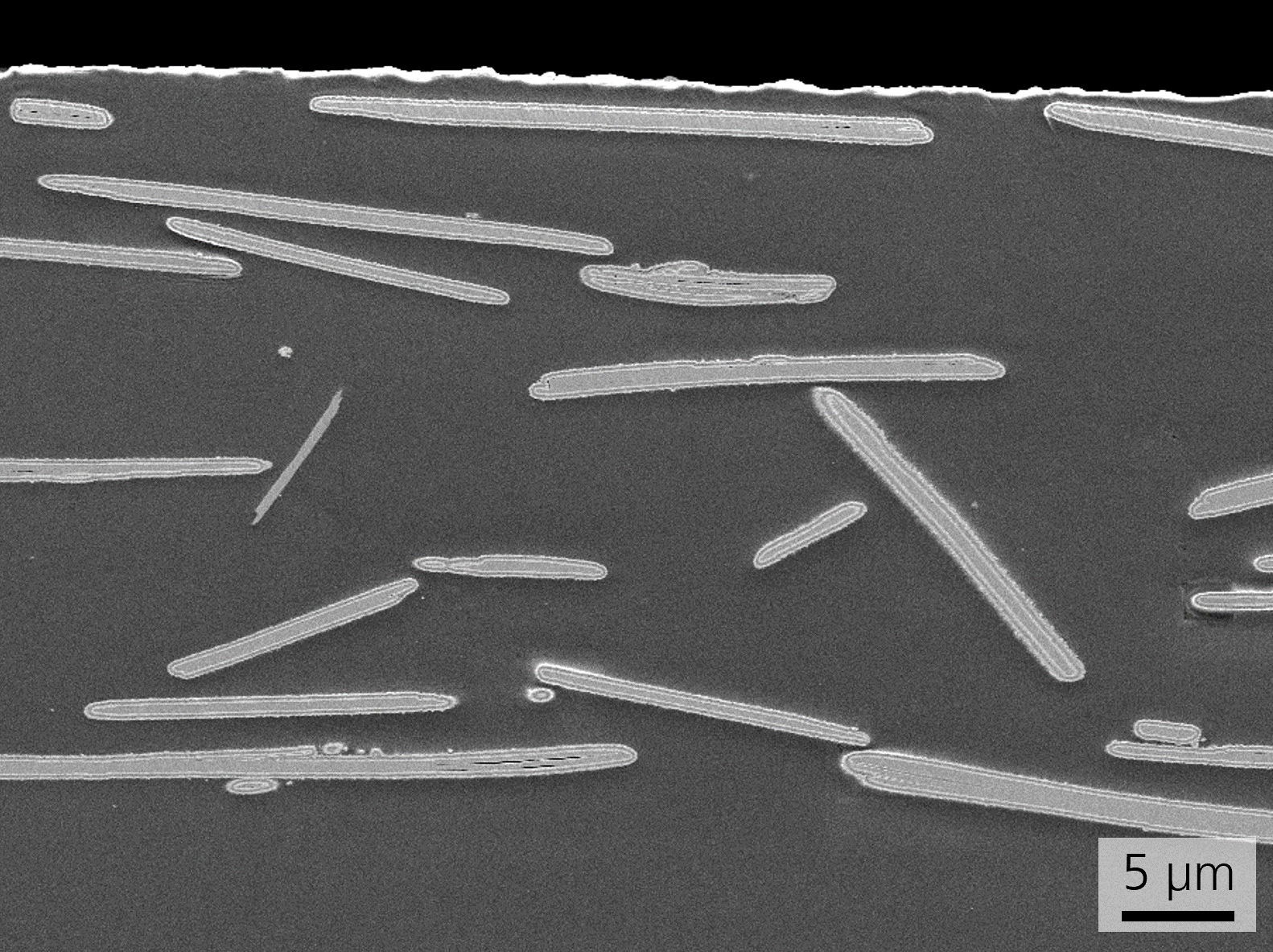
- How are the effect pigment particles distributed in the basecoat layer and what orientation do they have?
- What influence do microstructural parameters have on the macroscopic visual appearance of the coating system?
In addition to surface analytical techniques (such as time-of-flight secondary ion mass spectrometry), electron microscopic analysis methods provide the desired information.
The cross-sections required for this are often produced using temperature-controlled ion and laser beam-based cutting processes. In addition to individual pigments, entire coating systems can also be prepared without damage.
Scanning electron microscopy (SEM) on cross-sections generated using focused ion beams (FIB) is very well suited to imaging the embedding, position and orientation of the pigments in the coating system. Existing defects such as bubbles, holes or specks can thus be detected very well.
Transmission electron microscopy (TEM) is often used to gain deeper insights into the nanostructure of pigments. This enables the examination of the structural morphology, layer adhesion, interface properties or defect structure. Coatings with thicknesses < 10 nm are often used, which can only be analyzed using high-resolution TEM methods.
If a finely focused beam (STEM, beam diameter typically 1 nm) is used in the transmission electron microscope, even the finest details of the element distribution can be revealed.
The Fraunhofer IMWS has excellent equipment for microstructure diagnostics as well as decades of expertise in the characterization of various effect pigments. Given this background, we offer R&D services for the prompt contract analysis of coating systems and individual pigments.